In cardiac abnormalities with right-left shunts oxygen-poor blood gets from the right half of the heart into the left side and thus into the systemic circulatory system. In most of the defects with right-left shunts the physiologic shunts present before birth remain functional because no reversal of pressure happens following birth.
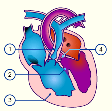
- RV outflow obstruction
- VSD
- RV Hypertrophy
- Overriding Aorta
II. Pulmonary valve atresia
This abnormality is very rare and accounts for only 1-3%. The pulmonary valve is atretic and there is no exit from the right ventricle. Thus, all of the blood flows into the left atrium via the foramen ovale and the lungs only get perfused via a very wide ductus arteriosus.
IIII. Tricuspid atresia
Incidence of 3%. Through atresia of the tricuspidal valve a connection between the right atrium and the right ventricle is missing. Since this abnormality is mostly combined with a VSD, its effects depend on the size of the VSD.
IV. Hypoplastic Left Heart Syndrome
A special case is the Hypoplastic left heart syndrome, a rare (only 2% of all cardiac abnormalities) that includes a poorly developed (hypoplastic) left ventricle that is often only a few millimeters thick. The aortic and mitral valves are constricted (stenotic) or completely closed (atretic). Only the right ventricle supplies both the pulmonary and systemic circulation system with blood. The arterial blood from the left atrium gets into the right atrium and ventricle via the patent foramen ovale (PFO) (L–>R shunt). The ascending aorta is filled retrograde by the blood of the ductus arteriosus (Rß L shunt). This way, the coronaries etc. are supplied with blood flowing retrograde via the PDA, the only way survival is possible for a short time.
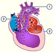
Hypoplastic Left Heart Syndrome
The left ventricle is hypoplastic. Through an open foramen ovale the blood gets to the right side and mixes with the O2 unsaturated venous blood. This left-right shunt via the foramen ovale as well as the right-left shunt via the ductus arteriosus are necessary for survival. Such children are cyanotic.
From:
Embryology
Posted on August 20, 2013 – 10:50pm
Cardiac defects without a shunt
1. Right Heart obstruction
Obstructions can occur at various levels in the right side of the heart or in the pulmonary outflow path. Pure obstructions do not result in a mixing of the blood but cause a massive additional strain on the ventricles because they have to pump against increased resistance.
Introduction to Cardiac congenital defects
Posted on November 26, 2010 – 8:36pm
Congenital cardiac disorders, with an incidence of 1/1000 newborns, belong to the most frequent birth defects. Chromosomal aberrations are frequently associated with heart abnormalities.
Before we go on, please review here the development of the heart:
Cardiac Defects with a Right to Left Shunt (Cyanotic)
In cardiac abnormalities with right-left shunts oxygen-poor blood gets from the right half of the heart into the left side and thus into the systemic circulatory system. In most of the defects with right-left shunts the physiologic shunts present before birth remain functional because no reversal of pressure happens following birth.
Cyanosis
Cyanosis is a blue coloration of the skin and mucous membranes due to the presence of greater than, or equal to, 2.5 g/dL of deoxygenated hemoglobin in blood vessels near the skin surface.
Syllabus of Clinical Thoracic and Cardiac Embryologic Problems with anatomic correlations
Posted on August 20, 2013 – 6:07pm
Case studies involving the thorax, mediastinum, embryology of the heart & circulatory systems
Anatomic and clinical reviews that will include basic symptoms and findings, diagnosis & treatment
Tufts University school of Medicine
Clinical Anatomy
Joan F. Tryzelaar, M.D., F.A.C.S, F.A.C.C.P.
January, 2011
Before we go on to congenital heart problems, it is good to review how to get into the chest:
Clinical Case:
You have just been invited to participate in an operation. The surgeon plans a posterolateral thoracotomy to enter the chest. He will tell you that he plans entry via the 6th intercostals interspace(Pearl: found two finger widths below the tip of the scapula).
Tetralogy of Fallot (TOF)
Tetralogy of Fallot, described in 1888, is seen in about 8% of all congenital cardiac abnormalities. It includes the following defects:
- Pulmonary stenosis (PS)
- Ventricle septum defect (VSD)
- Overriding aorta
- Right ventricular hypertrophy