Anatomy | Right Side | Left Side | Interior | Septum | Valves | Blood Flow
How your heart works
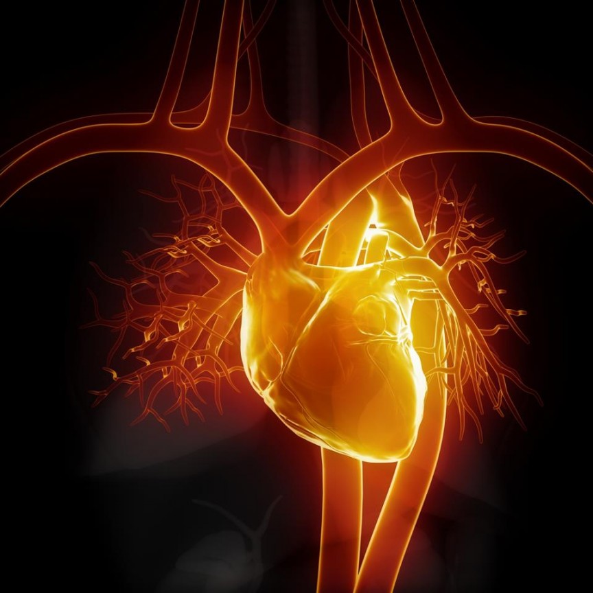
Your heart is located under the ribcage in the center of your chest between your right and left lungs.A normal, healthy, adult heart is about the size of an average fist:
Your heart is a muscle that pumps blood to the rest of your body. Your heart is part of your circulatory system, which consists of a network of arteries, veins, and capillaries that carry blood to and from all areas of your body. Electrical signals force your heart to contract and thus pump your blood to the rest of your body.
Your blood carries the oxygen and nutrients that your organs need to work normally. Blood also carries waste products such as carbon dioxide back to your heart and into your lungs to be passed out of your body and into the air. If disease or injury weakens your heart (your pump), your body’s organs won’t receive enough blood to work normally.
The heart has four chambers: The right and left atria and the right and left ventricles. The right side of your heart gets venous blood back from your body and then pumps it to your lungs. When you breathe in, oxygen passes from your lungs into your blood. Carbon dioxide, a waste product, is passed from the venous blood to your lungs and removed from your body when you breathe out.
The left atrium receives oxygen-rich blood from your lungs. The pumping action of your left ventricle sends this oxygen-rich blood to the rest of your body:
- Vena Cava
- Right Atrium
- Right Ventricle
- Main Pulmonary Artery
- Left & Right Pulmonary arteries
- Left & Right Pulmonary veins
- Left Atrium
- Left Ventricle
- Aorta
- Blood flow to the rest of your body
This animation shows how blood flows to and from your heart:
The Right Side of Your Heart
The superior and inferior venae cavae are in blue to the left of the heart muscle as you look at the picture. These veins are the largest veins in your body. After your body’s organs and tissues have used the oxygen in your blood, the venae cavae carry the venous (= oxygen-poor) blood back to the right atrium of your heart.
The superior vena cava carries venous blood from the upper parts of your body, including your head, chest, arms, and neck. The inferior vena cava carries venous blood from the lower parts of your body.
The blood from the venae cavae flows into your heart’s right atrium and then on to the right ventricle. From the right ventricle, the blood is pumped through the pulmonary arteries to your lungs. There, the blood picks up more oxygen.
The Left Side of Your Heart
The oxygen-rich (= arterial) blood passes from your lungs back to your heart, enters the left atrium and is pumped into the left ventricle. From there the arterial blood is pumped to the rest of your body through the aorta.
Like all of your organs, your heart needs blood rich with oxygen for its own energy supply.. This oxygen is supplied through the coronary arteries as blood is pumped out of your heart.
Your coronary arteries are initially located on your heart’s surface. Deeper branches eventually reach the muscle cells where the energy exchange takes place. Procedures such as Percutaneous Cardiac Interventions (PCI) and Coronary Artery Bypass Grafting (CABG) are only performed to the arteries on the surface. Your coronary arteries (shown in red in the drawing) carry arterial blood to all parts of your heart.
The Interior of Your Heart
Below is a picture of the inside of a heart:
The blue arrow shows the direction in which venous (oxygen-poor) blood flows from the body to the lungs. The red arrow shows the direction in which arterial blood flows from the lungs to the rest of the body (from: http://www.nhlbi.nih.gov/health/dci/Diseases/hhw/hhw_anatomy.html).
Your Septum
The right and left sides of your heart are divided by an internal wall of tissue called the septum (see above).
Your Heart Valves
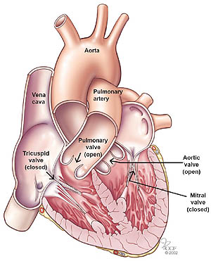
The picture shows your heart’s four valves. Their function is to direct your venous (oxygen-poor) blood flow forward to your lungs on the Right side from the right atrium to your lungs:
- Tricuspid valve, between the Right atrium and Right ventricle
- Pulmonary valve, between the Right ventricle and the Pulmonary artery
On the Left side oxygen rich blood from your lungs to your body:
- Mitral valve, between the Left atrium and Left ventricle
- Aortic valve, between the Left ventricle and aorta
Your Blood Flow
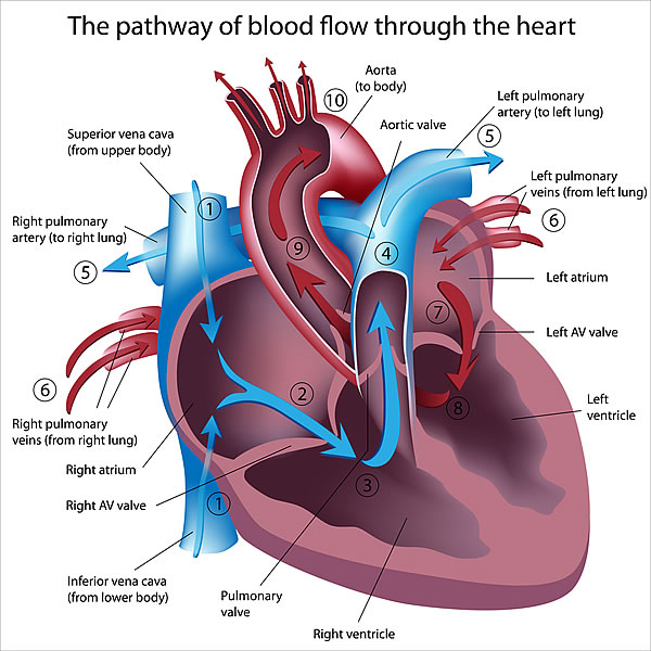
Once again, the arrows in the drawing show the direction that blood flows through your heart. The light blue arrows show that blood enters the right atrium of your heart from the superior and inferior venae cavae. From the right atrium, blood is pumped into the right ventricle.
From the right ventricle, blood is pumped to your lungs through the pulmonary arteries.The light red arrows show the arterial blood coming in from your lungs through the pulmonary veins into your heart’s left atrium. From the left atrium, the blood is pumped into the left ventricle. The left ventricle pumps the blood to the rest of your body through the aorta.
For the heart to work properly, your blood must flow in only one direction. Your heart’s valves make this possible. Both of your heart’s ventricles have an inlet valve from the atria and an outlet valve leading to your arteries.
Healthy valves open and close in very exact coordination with the pumping action of your heart’s atria and ventricles. Each valve has a set of flaps called leaflets or cusps that seal or open the valves. This allows pumped blood to pass through the chambers and into your arteries without backing up or flowing backward, just like a door closes a room.
(Adapted from National Heart Lung and Blood Institute, http://www.nhlbi.nih.gov/health/dci/Diseases/hhw/hhw_whatis.html)
Comments 2
what is left bundle branch block?
Do values slowly deteriorate? Does a heart murmur stabilize within the body’s ecosystem? Can valves with moderate regurgitation or leakage stabilize or do they get worse over time? What would be a progression towards a surgical intervention, and what would not require surgery? (I am 65 with 3 moderate regurgitation values.) Thank you.