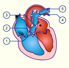
- Subvalvular (Right ventricular outflow tract)
- Valvular
- Supravalvular (pulmonary truncus)
- Peripheral (at the bifurcation)
- Peripheral in the pulmonary artery
Isolated PS occurs with a frequency of 9% of all congenital cardiac abnormalities and, combined with other cardiac abnormalities, with a frequency of nearly 21%.
All forms lead to an obstruction of blood flow from the right ventricle to the pulmonary arteries. Depending on how severe the stenosis is a compensatory hypertrophy of the right ventricle developes.
Pathophysiology:
Light to middle-degree pulmonary stenoses often do not show themselves clinically and the development of the child is normal. As a rule, larger stenoses lead to right ventricular hypertrophy and heart failure. Symptoms include:
- Dyspnea and tachypnea
- Hepatomegaly due to blood congestion
- Cyanosis may occur due to a persisting foramen ovale and R–>L shunt
Diagnosis:
In an ECG (signs of right hypertrophy), X-ray, ECHO.
Therapy:
Balloon dilatation or surgical correction.


Comments 1
Pingback: Cardiac defects without a shunt - Cardiac Health