
1. Patent Ductus Arteriosus (PDA)
Incidence: 9%
After birth, within the first days of life, the PDA is closed by contractions of its smooth muscle intima proliferation. This process is triggered by the postnatal pO2 increase with the first breath of the newborn.
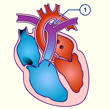
- In infants with a large PDA increasing cardiac insufficiency with tachypnea, dyspnea, and growth disorders are observed. Drinking weakness, accompanied by increased sweating occur when nursing because this represents a big effort for the newborn.
- In premature infants, an open PDA leads to a flooding of the pulmonary circulation system.
- In older children an open PDA is often discovered accidentally. It seldom leads to a symptom such as infection proneness.
2. Atrial Septal Defect (ASD)
Incidence: 11%
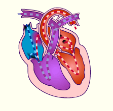
Right heart circulation system is overloaded because blood from the left side always recirculates through the lungs via the ASD.
3. Ventricular Septal Defect (VSD)
Incidence: (28%)
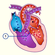 1 Defect in the membranous part of the inter ventricular septum
A VSD may occur by itself but is often associated with other defects.
After birth the severity of the VSD depends on its size and the resistance relationships in the pulmonary and systemic circulation systems.
4. Atrio-Ventricular Septal Defect (AV Canal)
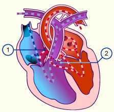
This cardiac abnormality occurs most frequently in patients with Down syndrome and is associated with a Defect in the atrial and Ventricular septum
From:
- Anatomy & Anomalies, Children’s Heart Center, Johns Hopkins University
- Heart Embryology, University of Toronto
- Cardiovascular Embryology, Dept. of Anatomy, Indiana University
- Human Embryoloogy, Organogenesis
Embryology
Posted on August 20, 2013 – 10:50pm
Cardiac defects without a shunt
1. Right Heart obstruction
Obstructions can occur at various levels in the right side of the heart or in the pulmonary outflow path. Pure obstructions do not result in a mixing of the blood but cause a massive additional strain on the ventricles because they have to pump against increased resistance.
Introduction to Cardiac congenital defects
Posted on November 26, 2010 – 8:36pm
Congenital cardiac disorders, with an incidence of 1/1000 newborns, belong to the most frequent birth defects. Chromosomal aberrations are frequently associated with heart abnormalities.
Before we go on, please review here the development of the heart:
Cardiac Defects with a Right to Left Shunt (Cyanotic)
In cardiac abnormalities with right-left shunts oxygen-poor blood gets from the right half of the heart into the left side and thus into the systemic circulatory system. In most of the defects with right-left shunts the physiologic shunts present before birth remain functional because no reversal of pressure happens following birth.
Cyanosis
Cyanosis is a blue coloration of the skin and mucous membranes due to the presence of greater than, or equal to, 2.5 g/dL of deoxygenated hemoglobin in blood vessels near the skin surface.
Syllabus of Clinical Thoracic and Cardiac Embryologic Problems with anatomic correlations
Posted on August 20, 2013 – 6:07pm
Case studies involving the thorax, mediastinum, embryology of the heart & circulatory systems
Anatomic and clinical reviews that will include basic symptoms and findings, diagnosis & treatment
Tufts University school of Medicine
Clinical Anatomy
Joan F. Tryzelaar, M.D., F.A.C.S, F.A.C.C.P.
January, 2011
Before we go on to congenital heart problems, it is good to review how to get into the chest:
Clinical Case:
You have just been invited to participate in an operation. The surgeon plans a posterolateral thoracotomy to enter the chest. He will tell you that he plans entry via the 6th intercostals interspace(Pearl: found two finger widths below the tip of the scapula).
PDA
PDA accounts for 5 to 10% of congenital heart anomalies; the male: female ratio is 1:3. PDA is very common among premature infants (in 45% with birth weight < 1750 g; in about 80% with birth weight < 1200 g). A significant PDA causes heart failure (HF) in 15% of premature infants with birth weight < 1750 g and in 40 to 50% of infants with birth weight < 1500 g.
Atrial Septal Defect
The Foramen Ovale in Fetal Circulation
Before birth, the fetal circulatory system includes three open structures through which blood moves that normally close soon after birth. These structures are the ductus arteriosus, the ductus venosus, and the foramen ovale. The foramen ovale allows the shunting of blood from the right atrium into the left atrium.
VSD
A Ventricular Septal Defect (VSD) is the second most common cardiac malformation, accounting for approximately one fifth of all congenital cardiac anomalies. VSD also occurs in combination with other congenital heart defects, as in an atrioventricular canal (AVC), tetralogy of Fallot (TOF) and occasionally, transposition of the great arteries (TGA).