- Introduction
- Stenosis, (valve too tight)
- Insufficiency (leaking valve)
- Mitral valve
- Aortic valve disease
- Complications of valve disease
- Valve disease and Coronary artery disease
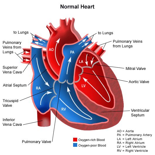
Your heart has four chambers: The right and left atria and the right and left ventricles, separated by four valves that control the flow of blood through your heart and the septum, which separates the left side from the the right side of your heart.
The four heart valves are:
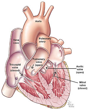
- The tricuspid valve, located between the right atrium and right ventricle;
- The pulmonary or pulmonic valve, between the right ventricle and the pulmonary artery;
- The mitral valve, between the left atrium and left ventricle; and
- The aortic valve, between the left ventricle and the aorta.
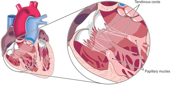
Each valve has a set of flaps (also called leaflets or cusps). The mitral valve has two leaflets; the others have three. Under normal conditions, the valves permit blood to flow in only one direction. Blood flow occurs only when there’s a difference in pressure across the valves that causes them to open. The mitral and tricuspid valves are connected to small muscles (papillary) along the wall of the heart by small string like tendons (chordeae tendineae). Papillary muscle contraction opens these valves.

The aortic and pulmonic valves are differently shaped and do not have cordae tendineae or papillary muscles.
Problems with a heart valve may occur because of disease, injury or congenital factors. When your heart valves become diseased or damaged, its leaflets can become thickened and rigid. When this occurs the valve may not be able to fully open or to completely close. This is called stenosis (too narrow) or insufficiency (too wide). This may cause blood to leak back or require the involved heart chamber to work harder to move blood across the narrowed valve. In time this condition will result in some form of heart failure if left untreated.
Valvular stenosis results from a narrowing of the valve orifice that is usually caused by a thickening and increased rigidity of the valve leaflets, often accompanied by calcification. When this occurs, it results in obstruction of flow at the level of the valve involved.
Valvular insufficiency results from the valve leaflets not completely sealing when the valve is closed so that a backward flow of blood (regurgitation) of blood occurs into the heart. Mitral Valve Prolapse (MVP) and Bicuspid Aortic Valve disease (BAV) are examples of leaky valve diseases.

What are the symptoms of a leaking mitral valve?
Many patients with mitral valve disease are have no symptoms, even with a leak that is severe. When symptoms develop, they include shortness of breath, fatigue, loss of energy, swelling of the ankles and palpitations (extra or skipped heart beats).
How is a leaky mitral valve diagnosed?
The first step involves listening with a stethoscope. Using a stethoscope, the doctor hears a murmur, which represents turbulent blood flow across an abnormal valve. The diagnosis is confirmed by an echocardiogram.
Papillary muscle dysfunction ( papillary muscles do not work properly) may occur from a heart attack or cardiomyopathy and heart failure. This can cause regurgitation to occur across the tricuspid or mitral valves. Rupture of a papillary muscle (usually after a heart attack) may cause sudden regurgitation of blood back into the lungs. This may cause severe breathing problems due to excess fluid in the lungs- this is called congestive heart failure.
What is mitral valve prolapse?
Mitral valve prolapse is a common condition in which the mitral valve leaflets are floppy or loose.
Aortic valve disease occurs when the aortic valve does not work correctly. This can be caused by:
- Aortic valve stenosis: Stiff, fused, inflexible valve leaflets that lead to the narrowing of the aortic valve, which limits or blocks the blood flow. Aortic valve stenosis occurs when calcium is deposited on the valve leaflets, limiting their mobility. Stenosis can occur in patients with a normal (3 leaflets) or a bicuspid (2 leaflets) aortic valve.
- Aortic valve regurgitation (also called valvular insufficiency, incompetence or “leaky valve”): Valve leaflets that do not close completely. Regurgitation causes the valve to leak, which limits the forward flow of blood through the aortic valve. Regurgitation may occur because of floppy leaflets (prolapse), infection of the valve (endocarditis), dilatation of the aorta (aneurysm), and rheumatic valve disease.
The aortic valve may be abnormal at birth (congenital aortic valve disease) or become diseased over time (acquired valve disease). With acquired aortic valve conditions, changes occur in the structure of the valve. Acquired aortic valve conditions include:
- Infective endocarditis is a bacterial infection of the valve, which is caused when bacteria enter your blood stream from the site of a remote infection and attach to the surface of your heart valves. Even minor infection, such as a tooth abscess can cause severe bacterial endocarditis of the aortic valve.
- Rheumatic fever is usually caused by a bacterial throat infection, such as strep throat. The valve itself is not infected in rheumatic fever, but antibodies developed by the body to fight infection react with the heart valves, causing stiffening and fusion of the leaflets of the aortic valve.
- Aortic valve degeneration is another cause of acquired aortic valve disease. In many patients, the aortic valve leaflets degenerate and become calcified with time. This most frequently causes aortic stenosis, but may also cause aortic regurgitation. This is the most common cause of aortic stenosis in people over the age of 65.
- Other causes of aortic valve disease include: heart attacks, syphilis, hypertension, aortic aneurysms, connective tissue diseases, and less commonly, tumors, some types of drugs and radiation.
Both stenosis and insufficiency can have serious cardiac consequences, and produce the following symptoms:
- Shortness of breath (dyspnea)
- Fatigue
- Reduced exercise capacity
- Light headedness or fainting (syncope)
- Heart failure
- Pulmonary hypertension
- Pulmonary/systemic edema
- Chest pain (angina)
- Arrhythmias
- Blood clots (thromboembolism) which can cause stroke
Valvular problems may be caused by infection, heart disease, trauma or congenital valvular conditions and may be isolated to a single valve or effect multiple valves. Right sided (tricuspid, pulmonary) valvular disease is much less common than left sided (aortic, mitral) valvular disease. Roughly 90% of valvular disease is chronic, having developed gradually over many years. Complications of rheumatic fever, congenital disorders and aging cause the vast majority of chronic valvular disease. The remaining 10% of valvular disease that develops acutely (over days to weeks) is often due to complications of recent heart attack or infections.
Coronary Artery Disease associated with a heart valve problem
If you are diagnosed with a valve problem in addition to coronary artery disease, the benefits of surgery are often even more significant. However, it generally also increases the risk of an operation, particularly if there has been a lot of damage to your heart.
Unlike Coronary artery bypass grafting (CABG), valve surgery encompasses a much more diverse group of operations. Since there are four cardiac valves, they may malfunction in a number of different ways. The valves may be repaired or replaced with a wide range of techniques. Sometimes, multiple valves in combination with CABG may need to be repaired or replaced.
Since many different procedures may need to be considered in a patient with CAD and an additional valve problem, it is in general difficult to provide an easy answer. On the other hand, a new valve, particularly as replacement of an Aortic valve, or repair of a leaking Mitral valve can often lead to amazing improvement in the quality of your life, thus making it well worth taking a risk for. Here it is particularly important to have an experienced surgical team take care of you!
Comments 1
Since my tricuspid and mitral valve replacement in 2018 I have over the last year become darker in my skin colour is this because of blood circulation ? I have also developed gouty arthritis in my fingers and toes .I am 79 …and still active.