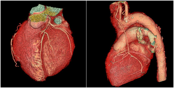
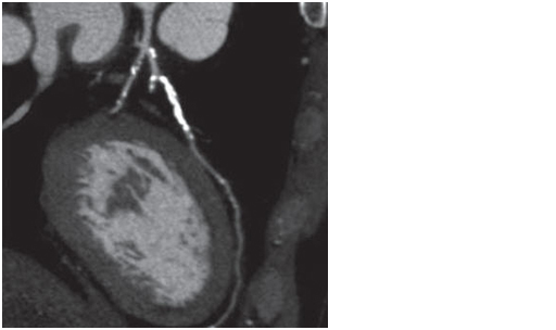
Coronary CT angiography (CCTA) is a noninvasive imaging modality which can be used to evaluate the anatomy of the coronary arteries. CCTA allows direct visualization of the coronary artery wall and lumen with the administration of intravenous contrast. The degree of coronary luminal stenosis can be reliably estimated, as can the presence or absence of both calcified and non-calcified plaques.
Conventionally, non-invasive methods for detection of coronary disease using stress treadmill testing, stress echocardiography and myocardial perfusion scan provide indirect means of diagnosing significant coronary disease where the coronary luminal diameter narrowing is more than 70 per cent. But they are unable to detect the vulnerable plaques that do not cause any significant functional stenoses, though they account for the large number of patients who eventually develop ACS. Coronary CT Angiography (CCTA) is one of the new modalities of coronary artery imaging. It is playing an important role in the assessment of coronary artery disease.
CCTA can be an effective diagnostic tool for select patients presenting to the emergency room with acute chest pain. CCTA, with its 100% negative predictive value, can virtually eliminate the elective negative diagnostic coronary catheterization in low- and intermediate-risk patients (i.e. those with negative cardiac enzymes and no ECG changes). Some studies have shown that the use of CCTA may decrease hospital admission time as well as improve cost effectiveness compared to traditional diagnostic strategies used to evaluate acute chest pain. The American College of Cardiology (ACC) appropriateness guidelines support the use of CCTA in intermediate-risk patients presenting with acute chest pain. One of the more common uses of CCTA in current clinical practice is to evaluate for CAD in patients with “equivocal” or “non-diagnostic” stress test results.
The medical literature has shown that CCTA compares favorably against stress tests as an initial exam and against catheter coronary angiography for examining obstructive coronary artery disease. The cost savings of using a protocol that incorporates early use of CCTA in emergency and nonemergency settings for low- to intermediate-risk coronary artery disease patients has been well documented.
Safety
Radiation exposure
Prospectively gated CCTA is now performed with a dose range of 0.4 to 10 mSv, which is well below the SPECT stress test range. With a sestamibi-sestamibi protocol, radiation doses range from 12 to 25 mSv. When thallium is incorporated into the protocol, as either a single or dual isotope, radiation doses can easily exceed 22 to 30 mSv per test.
Contrast
Contrast risks are low and comparable for both CCTA and Coronary angiography for native coronary angiography. In the case of ICA of bypass grafts, the contrast load will be significantly higher as compared to CCTA. With the 64 slice CT, Coronary CTA can be performed with as low as 50cc of iodinated contrast. The precautions to be taken for those with renal insufficiency are the same as in any examination, which require contrast agents.
CCTA can provide three dimensional views of the coronary vessel wall structure, heart muscle, valves, pericardium and even ventricular contractility function. It has a higher sensitivity and specificity for the detection of coronary artery disease compared with conventional non-invasive tests. CCTA can provide a three dimensional view of the coronary vessel wall structure, heart muscle, valves, pericardium and even ventricular contractility function. It has a higher sensitivity and specificity for the detection of coronary artery disease compared with conventional non-invasive tests.
The advent of 256 and 320-slice CT scanners will eliminate many of the technical difficulties that affect the temporal resolution of coronary CT angiogram, presently requiring a patient holding his breath for 7 to 10 seconds with the 64-slice CT scanner. With newer 256 and 320-slice CT scanners the scanning time will be about 1 to 2 seconds.. As technology advances, MDCT imaging of the coronary arteries will become the diagnostic tool of choice for the detection of coronary artery disease.