Transposition of the great arteries (TGA) is a life-threatening malformation in neonates.
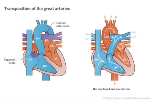
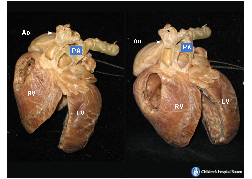
While the cause of TGA (5%) is unknown, an increased risk is associated with maternal diabetes mellitus. The great arteries are parallel at the base of the heart. The aorta (Ao) is anterior to the pulmonary artery (PA) and arises from the right ventricle (RV). The pulmonary artery arises from the left ventricle (LV). From an embryologic point of view, the rotation of the aorto-pulmonary septum of 180⁰ did not take place.
The hemodynamics of TGA are characterized by a parallel connection of the two circulatory systems. This situation can only be survived when mixing of O2 via a shunt can take place:
- Atrial level: Patent Foramen Ovale (PFO) or Atrial Septal Defect (ASD),
- Ventricuar level: Ventricular Septal Defect (VSD),
- Systemicl level: Patent Ductus Arteriosus (PDA).
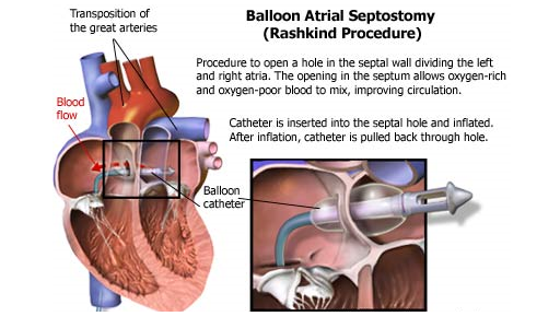
At least one-third of newborns with TGA will require urgent intervention within hours of birth, usually a procedure to allow mixing of the two separate (parallel) circulations via the creation (or widening) of the Foramen Ovale via what is called an Atrial Septostomy.
All babies have a foramen ovale between the left and right atrium in utero which closes spontaneously after birth. In some heart problems (eg. transposition, tricuspid atresia) it is often necessary for foramen ovale to remain open for the blood to circulate through the heart. If this is the case and the natural hole is too small then the hole is enlarged via a Balloon Atrial Septostomy:
Final Treatment is via surgical correction: