Sometimes when cardiologists read EKGs the results will show a right bundle branch block, left bundle branch block or incomplete bundle branch block. This term refers to a particular appearance of the QRS complex on the EKG. The EKG represents the electrical activity of the heart and we use it to look not only at the heart rate and rhythm but also we can determine certain characteristics of the electrical system of the chambers the heart.
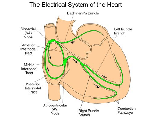
The electrical system of the heart is composed of three major components. The first is the SA node, which is a group of cells close to the top of the heart where the superior vena cava joins the right atrium. The second is the AV node. The third component is called the Purkinje fibers or Purkinje bundle. There is one Purkinje bundle for the right ventricle and two Purkinje bundles for the left ventricle. These bundles are connected with the AV node by what is called the bundle of His.
Normally when the heart begins to contract electrical activity originates in the SA node. This causes a wave of electrical impulse to go through the muscle tissue of the two atria of the heart or upper chambers. This causes the upper chambers to contract and is seen on the EKG as the P wave. Normally this electrical activity generated by the SA node can pass to the ventricles is through the AV node. The AV node acts as what could be considered a delay switch to allow time for blood to pass from the atria down into the ventricles and fill the ventricles. This delay can be measured by what we call on the EKG as the PR interval. Once the electrical impulse has passed through the AV node, the impulse is rapidly transmitted across the bundle of His to the Purkinje bundle systems down to the bottom (apex) of the ventricles. This stimulates the ventricles to contract simultaneously.
As the ventricles contract, the blood is ejected, either toward the pulmonary artery and out to the lungs or out the aorta distributed to the body. The contraction of the ventricles is demonstrated on the EKG by what is called the QRS complex. After the ventricles have contracted and the electrical impulses given off, the heart muscle then recharges to be ready for the next contraction. This period of time can be seen on the EKG by what is called the T wave.
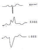
Bundle branch block, either complete or incomplete, represents a difference in the timing of the arrival of the electrical impulse at the apex of ventricle and can be caused by many different reasons. One of the causes of bundle branch block either right or left can be the surgical interruption of the Purkinje bundle. For example, when a patient has had heart surgery where the right bundle has been cut or damaged and the left bundle is intact this condition is known as right bundle branch block (RBBB). In this case the impulse initiates the contraction of the left ventricle with a delay of the contraction of the right ventricle. This does not mean that the right ventricle does not contract but merely that there is a delay in the contraction process of the right ventricle. Likewise, there are times where there can be a left bundle branch block (LBBB) caused by surgical interruption of the left-sided bundles and this again signifies that the right ventricle contracts first and then the left ventricle contracts after that, but again does not mean that the left ventricle does not contract at all. We sometimes can see bundle branch block patterns with enlargement of either the left or right ventricle where the Purkinje fibers are stretched. This stretching causes a slight delay in the contraction of the ventricle that is enlarged.
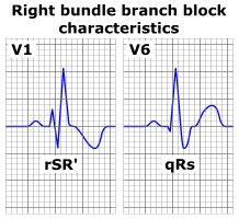
One sometimes will see an interpretation of an EKG described as either an incomplete right bundle branch or incomplete left bundle branch block pattern. The interpretation of incomplete or complete bundle branch block is determined by how wide the QRS complex is when measured over time. If the QRS complex is wider than 0.12 seconds that is considered a complete right bundle or left bundle branch block.
When we see patients that have these types of patterns on the EKG, usually a physical exam is adequate to determine whether or not this represents an abnormality of the heart. Sometimes it is necessary to obtain an echocardiogram to look at the cardiac structures. The term bundle branch block is not related to another term used with EKG interpretation called AV block. AV block represents a potential problem with the AV node.
Left bundle branch block may be associated with:
Left bundle branch block (LBBB) is a common electrocardiographic (ECG) abnormality seen in patients whose normal cardiac conduction. Although LBBB is often associated with significant heart disease and is often the result of myocardial injury, strain or hypertrophy, it can also be seen in patients without any particular clinical disease. However, LBBB can an important finding, especially in patients presenting with acute chest pain, syncope and in those suffering from heart failure with reduced ejection fraction.
Right bundle branch block may be associated with:
-
A heart abnormality that’s present at birth (congenital) — such as atrial septal defect, a hole in the wall separating the upper chambers of the heart
-
A viral or bacterial infection of the heart muscle (myocarditis)
-
High blood pressure (hypertension)
-
Scar tissue that develops after heart surgery
The complications of a bundle branch block are similar whether the blockage is on the right or left side of your heart. Complications may include :
-
Slow heart rate (bradycardia). Some of these patients may need to have a pacemaker.
-
Cardiac arrest or sudden cardiac death
People who have a heart attack and develop a bundle branch block have a higher chance of complications and death than do people who have heart attacks and don’t develop a bundle branch block.
Incomplete Right Bundle Branch Block
If there is a pattern similar to a bundle branch block but the width of the QRS complex is less than 0.12 seconds, we consider that to be an incomplete bundle branch block pattern. Incomplete right bundle branch block is very commonly seen and can be considered a normal variant of the QRS complex. This is frequently seen in teenagers and does not necessarily represent a serious heart condition.
Recommendation:
Incomplete RBBB does not require further evaluation in the presence of a negative family/personal history and physical examination. Because incomplete RBBB is a typical ECG finding in patients with an ‘ostium secundum’ atrial septal defect, symptoms and a fixed split of the second heart sound on auscultation should be excluded.
Bundle Branch Block in athletes
Physiological ECG abnormalities are more prevalent and significant in male athletes and athletes of African/Caribbean descent, one of which (even if quite rare) is mentioned here:
The Brugada syndrome is a hereditary disease that is associated with high risk of sudden cardiac death. It is characterized by typical ECG abnormalities: ST segment elevation in the pre-cordial leads (V1 – V3).
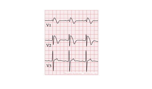
Typical ECG abnormalities in Brugada syndrome: ST elevation in V1-V3, without ischemia.
Comments 1
Have incomplete Right bunch block as per ECG.ECG taken due to dizziness near faint stage, fatigue, retching sensation, chest pain. Am on medication Calaptin 40 -3 times for AFib when pulse shot up last Sept to 160.